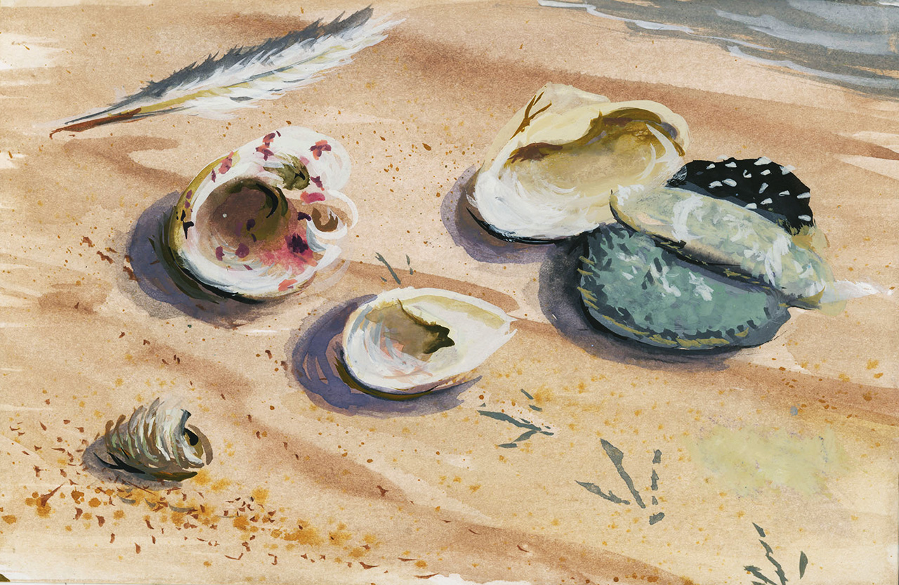
ANNUAL EXHIBITION 2021
OYSTERS UNDER THE MICROSCOPE
Photos and Videos by Steve Malinowski,
Fishers Island Oyster Farm
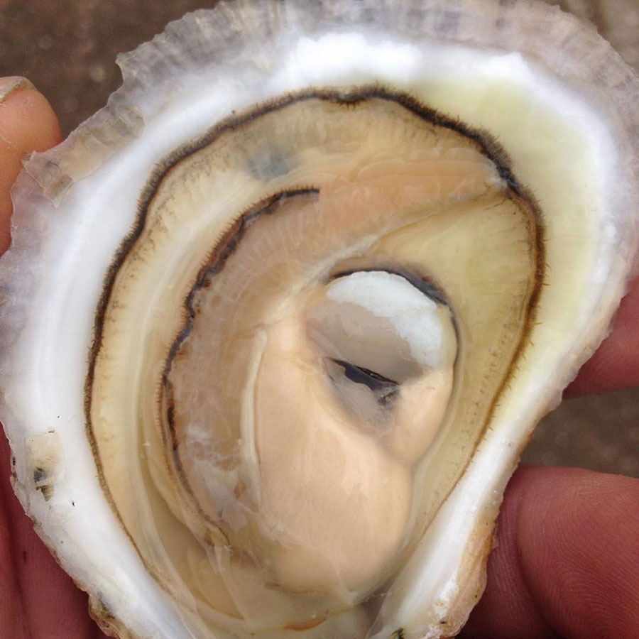
Ripe oyster showing gonad full of eggs
Photograph by Steve Malinowski

Set up for spawning oysters
Photograph by Steve Malinowski
Female oysters releasing eggs – millions at a time
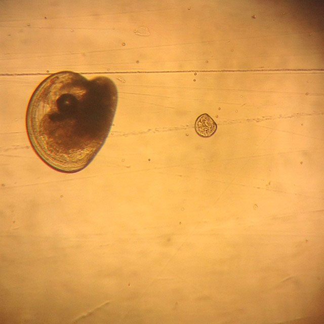
Fertilized oyster eggs prior to cell division
Photograph by Steve Malinowski
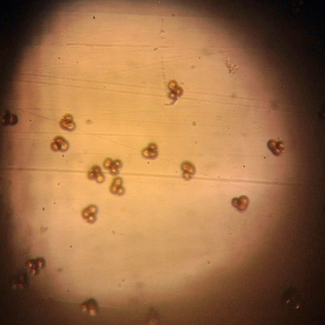
Fertilized oyster eggs showing first cell division 45 minutes after fertilization
Photograph by Steve Malinowski
Trochophores swimming four hours after spawning.
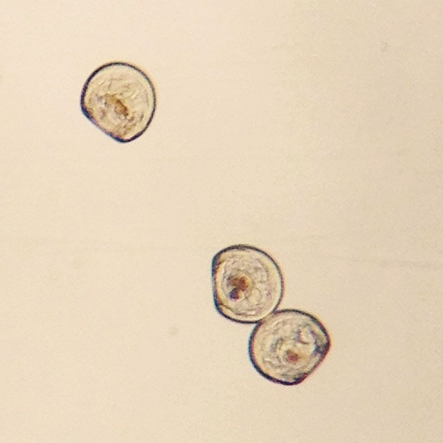
Two day old oyster larvae with cultured phytoplankton in gut. 80 microns
Photograph by Steve Malinowski
Phytoplankton culture system
Food under microscope
Two day old oyster larvae with cultured phytoplankton in gut. 80 microns
Photograph by Steve Malinowski

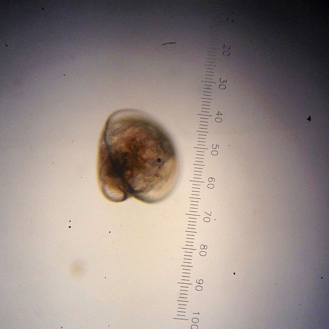
Eyed oyster larvae ready to go through metamorphosis. 300 microns.
Photograph by Steve Malinowski
Oyster larvae swimming with ciliated velum
Oyster larvae swimming and crawling about ready to set.

Newly set oyster having gone through metamorphosis
Photograph by Steve Malinowski
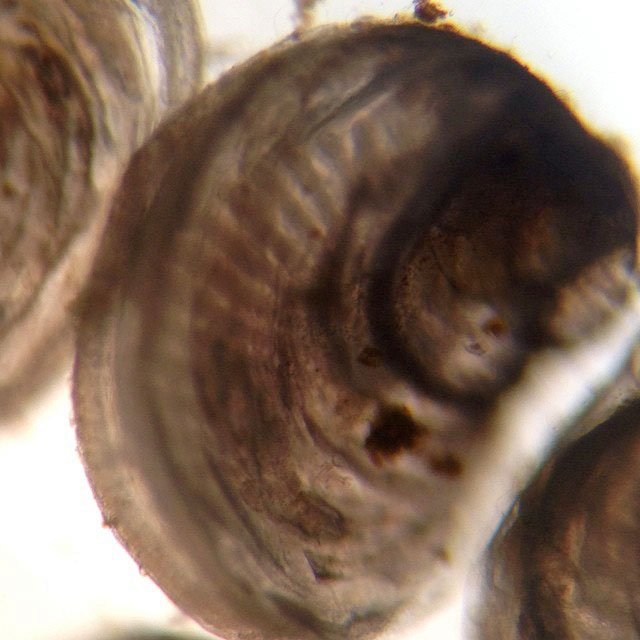
Newly set oyster with gills visible through shell, fingerlike structures.
Photograph by Steve Malinowski
Cilia pumping water and collecting food.
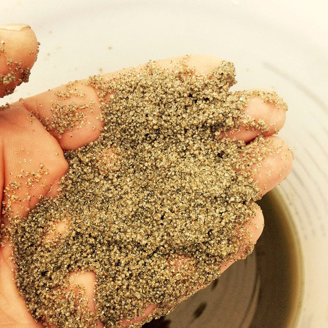
First Screening of seed after metamorphosis. 500-750 microns.
Photograph by Steve Malinowski
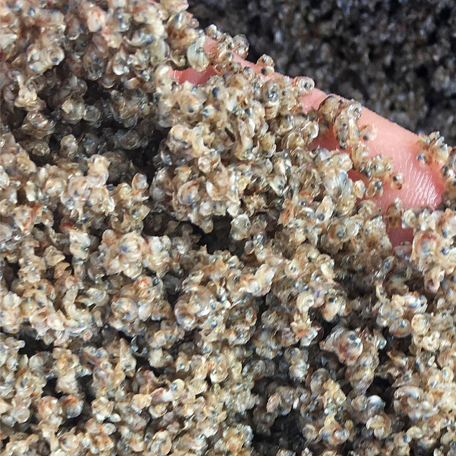
Seed ready to leave the factory.
Photograph by Steve Malinowski
Annual exhibition sponsored by ALTUS Partners & CHUBB